


You are resting on the living room rug with your senior dog, running your hands along their ribs or belly during a quiet evening petting session. Suddenly, your fingertips slide over a soft, squishy dome beneath the skin.
Your stomach drops. Finding any unexpected mass on an aging companion immediately triggers worry about canine cancer. You gently press the area, noticing that the lump moves freely under the coat and feels almost like a pocket of soft dough.
In most older dogs, this soft subcutaneous mass is a dog lipoma. While these fatty deposits are extraordinarily common and non-cancerous, assuming every lump is safe without veterinary confirmation carries real risks.
Quick Answer:
A dog lipoma is a benign, non-cancerous tumor composed of mature fat cells that commonly develops under the skin of senior dogs aged seven and older. Lipomas feel soft, rounded, and movable. They do not spread to other organs and rarely require surgery unless their size or location interferes with your dog’s walking, breathing, or daily comfort. A veterinary fine needle aspirate (FNA) is essential to confirm the diagnosis and rule out look-alike malignancies.
Key Takeaways:
- Benign Nature: Simple lipomas are harmless fatty collections that will not metastasize to internal organs or shorten your dog’s lifespan.
- In-Clinic Testing: A painless fine needle aspirate (FNA) collects microscopic fat droplets, confirming the lump is not a mast cell tumor or sarcoma.
- Selective Surgery: Removal is only necessary if the mass sits in high-friction areas like the armpit or groin, or causes physical pain.
- Lump Mapping: Measuring each bump monthly with a ruler helps your vet catch sudden texture changes or accelerated growth early.
What Is a Dog Lipoma? The Biology of Fatty Tumors
A lipoma is a benign neoplasm that arises from adipocytes, the specialized cells responsible for storing fat in canine body tissue. As dogs advance into their senior years, their cellular turnover slows down, and clusters of these fat cells occasionally overgrow within the subcutaneous layer between the skin and underlying muscle.
These cells stay enclosed within a thin fibrous capsule. Because the capsule isolates the fatty tissue, a standard lipoma does not invade surrounding muscles or send dangerous secondary cells into the bloodstream.
Veterinary oncologists see lipomas in canines of every breed. However, retrievers, cocker spaniels, dachshunds, and overweight senior dogs experience the highest rates of fatty lump development. Genetics and body condition both influence how many lipomas an older dog forms over time.
If you also notice small cauliflower-like blemishes on the surface of your companion’s skin, compare them with our guide on dog wart causes and removal in senior dogs.
Simple Lipomas, Infiltrative Lipomas, and Dangerous Look-Alikes
Not every fatty mass behaves the same way under the skin. Veterinarians classify fatty tumors into distinct categories based on how their cells interact with adjacent tissue.
The vast majority of lumps are simple lipomas. These sit loosely in the subcutaneous fat, move smoothly when gently pushed, and grow very slowly over months or years. They cause zero systemic illness and rarely cause pain.
A less common variant is the infiltrative lipoma. While still technically non-cancerous, infiltrative lipomas lack a tight fibrous border. They send microscopic tendrils of fat into surrounding muscle bellies, tendons, and connective tissue, making them feel firmer and firmly anchored in place.
The greatest concern for pet owners is distinguishing a harmless fatty lump from a malignant soft tissue tumor. Mast cell tumors, known among clinicians as the great imitators, often feel soft and doughy on initial palpation. A cancerous liposarcoma or fibrosarcoma can also initially hide beneath loose skin.
According to surgical guidelines from the American College of Veterinary Surgeons, accurate tissue identification before surgical intervention is crucial for planning clean margins and protecting muscle function.
| Mass Type | Texture & Feel | Mobility | Growth Rate | Diagnostic Confirmation |
|---|---|---|---|---|
| Simple Lipoma | Soft, doughy, smooth borders | Moves freely under the skin | Slow (months to years) | Fine Needle Aspirate (FNA) |
| Infiltrative Lipoma | Firm, deep, poorly defined edges | Fixed to underlying muscle | Moderate to slow growth | Biopsy & CT or MRI imaging |
| Mast Cell Tumor | Variable (soft, rubbery, or firm) | Can be movable or attached | Rapid or fluctuates in size | FNA cytology (granules) |
| Liposarcoma | Firm, nodular, irregular shape | Fixed to deep body structures | Rapid, aggressive progression | Histopathology tissue biopsy |
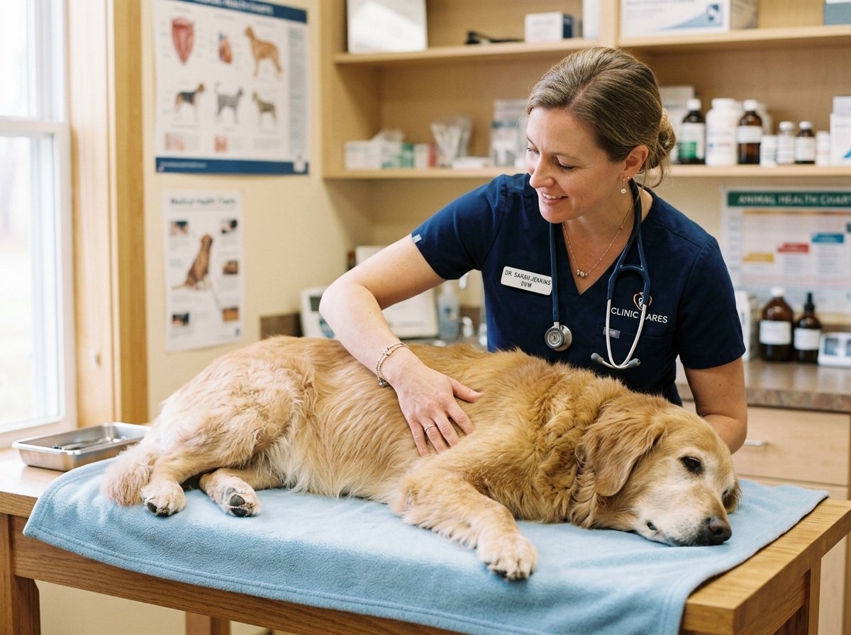
Why You Cannot Diagnose a Dog Lipoma by Touch Alone
Many pet owners make the dangerous mistake of assuming that any soft, movable lump is automatically harmless. Experienced veterinarians will tell you that even forty years in clinical practice cannot replace laboratory testing.
A soft texture merely indicates fluid or adipose density. Mast cell tumors frequently produce swelling that feels identical to a fatty lipoma. Without checking the cellular contents, you cannot know what lives inside that mass.
Fortunately, getting a definitive answer is fast, simple, and affordable. Veterinarians use a routine in-clinic procedure called Fine Needle Aspiration, commonly abbreviated as an FNA.
During an FNA, your veterinarian gently stabilizes the lump and inserts a very fine, sterile hypodermic needle. The needle collects a minute cluster of cells from within the center of the mass without requiring sedation or general anesthesia.
The sample is expelled onto a clean glass microscope slide, stained with laboratory dyes, and evaluated right in the clinic. If the lump is indeed a benign lipoma, the slide shows large, clear lipid droplets and mature adipocytes with small, quiet nuclei.
Research published by the Cornell University College of Veterinary Medicine highlights that early cytology remains the safest, most cost-effective first step for evaluating any new canine skin mass.
To learn more about tracking diverse lumps across your pet’s body, review our guide on old dog skin bumps and lumps.
When Does a Dog Lipoma Require Surgical Removal?
Finding out that your dog has a lipoma is often an immense relief. Once the needle aspirate confirms the benign diagnosis, the next question is if you should have the lump surgically cut out.
In the vast majority of cases, the answer is no. Surgery is not required simply because a lipoma exists. Because these tumors are completely non-cancerous, leaving them alone does not jeopardize your dog’s long-term health.
However, there are specific functional situations where veterinary surgery becomes necessary. Location matters far more than the simple diagnosis.
1. Mobility and Gait Interference
A lipoma positioned in the axillary space (the armpit) or the inguinal region (the inner groin) can cause mechanical difficulty. As the mass expands, it rubs against the moving leg with every stride.
Over time, this constant friction leads to muscle strain, joint compensation, and painful chafing. If a mass alters your dog’s natural walking rhythm or prevents them from climbing stairs, surgical removal restores comfortable mobility.
2. Rapid Expansion and Extreme Size
While most lipomas grow at a snail’s pace, some develop an extensive vascular network and grow into massive weights. A five-pound lipoma hanging from the chest or abdomen puts immense strain on an older dog’s spine and abdominal wall.
Removing these giant masses before they stretch the skin to its absolute limit prevents complex reconstructive surgery later. Earlier intervention on large masses is much simpler than waiting until the lump reaches extreme dimensions.
3. Skin Ulceration and Discomfort
When a lipoma grows very large, the overlying skin becomes thin and fragile. The skin can crack, bleed, or develop secondary bacterial infections from rubbing against carpets or bedding.
If your senior companion begins chewing, licking, or scratching at the lump, the mass is causing active irritation. If you suspect your dog is dealing with physical distress, check our reference on identifying senior dog pain signs.
Senior Dog Anesthesia Safety: Weighing the Risks
Deciding on surgery for a dog aged ten, twelve, or fourteen brings understandable anxiety about general anesthesia. Many owners fear their older dog will not wake up from the procedure.
Old age is not a medical disease. Advanced age simply means your dog has less physiological buffer for sudden blood pressure drops or organ strain. Modern veterinary medicine approaches senior anesthesia with rigorous safety protocols.
Before any surgery is scheduled, your vet will perform a detailed senior blood panel. This checks kidney values (BUN, creatinine, SDMA) and liver enzymes (ALT, ALP) to confirm the body can metabolize anesthetic agents smoothly.
Chest radiographs and an electrocardiogram (ECG) are often recommended to screen for hidden heart disease. During the procedure, your dog receives intravenous fluids, constant warming blankets, and dedicated monitoring of oxygen levels, blood pressure, and carbon dioxide output.
If a lipoma is small, harmless, and located on the back or flank, the anesthesia risk outweighs any cosmetic benefit. However, if the lump severely compromises daily walking, modern surgical care is exceptionally safe and highly beneficial.
Clinical data compiled in the Merck Veterinary Manual confirms that individualized senior anesthesia protocols reduce surgical risks to levels nearly matching younger patients.
At-Home Monitoring and Creating a Canine Lump Map
If your veterinarian confirms that your dog’s lipoma is safe to leave alone, your primary job becomes active at-home monitoring. Relying on memory to track changes in size or firmness is notoriously unreliable.
Veterinary clinics recommend creating a canine lump map in a small notebook or on your smartphone. Draw a simple outline of a dog, marking the exact location of each confirmed mass.
Once every month during a gentle grooming session, measure each lump across its widest point using a flexible fabric tape measure or plastic calipers. Record the date, the size in millimeters or centimeters, and notes on how the lump feels.
Healthy lipomas change very little from month to month. If you feel a lump suddenly double in size over three weeks, or if a previously soft lump becomes rock-hard and fixed to the ribs, contact your veterinarian for a recheck aspirate immediately.
Can Diet, Exercise, or Supplements Shrink a Dog Lipoma?
The internet is full of home remedies claiming that apple cider vinegar, herbal compresses, or special teas can dissolve canine lipomas. Pet owners naturally want a natural solution that avoids surgery.
Unfortunately, biological science tells a very different story. A lipoma is not general body fat that shrinks when calories drop. It is a distinct, encapsulated cellular tumor with its own independent microvascular blood supply.
Even if an overweight dog goes on a strict diet and loses five pounds, their lipomas will usually remain the exact same size. No dietary supplement, topical cream, or homeopathic herb has ever been proven to dissolve an established lipoma.
However, maintaining a lean body condition is still critically important for your senior dog’s everyday vitality. Excess body weight increases systemic inflammation and puts devastating stress on arthritic hips and knees.
Feeding a nutrient-rich, portion-controlled diet helps prevent your dog from developing additional fatty deposits. For healthy dietary transitions, explore our guide on selecting weight management dog food for older dogs.
Frequently Asked Questions
Can a dog lipoma turn into cancer?
No, a true simple dog lipoma will not transform into a malignant cancer over time. Lipomas are composed of mature, genetically stable fat cells that stay benign throughout your dog’s life. However, a malignant tumor like a liposarcoma or mast cell tumor can develop right next to an existing lipoma, which is why having every newly discovered lump aspirated by your veterinarian is vital.
How much does it cost to remove a lipoma on an older dog?
The cost to surgically remove a dog lipoma ranges from five hundred dollars to fifteen hundred dollars at most general practice veterinary clinics. Complex removals involving large infiltrative masses in the armpit or groin performed by a board-certified veterinary surgeon can cost two thousand dollars or more. The total price includes pre-anesthetic bloodwork, IV fluids, anesthesia monitoring, surgical time, pain medications, and post-operative pathology.
Does losing weight make a dog’s lipoma go away?
No, weight loss will not eliminate or shrink an established dog lipoma. Because lipomas are encased in an independent fibrous capsule with dedicated blood vessels, the body does not metabolize their stored lipids during caloric restriction. While shedding excess weight improves your senior dog’s joint comfort and stamina, the lipoma itself will retain its physical mass.
Can a dog have multiple lipomas at the same time?
Yes, many senior dogs develop multiple lipomas across different areas of their body as they age. Having three, five, or even ten separate fatty deposits on the chest, belly, and thighs is very common, especially in breeds like Golden Retrievers, Labradors, and Weimaraners. Each individual lump must be aspirated separately to confirm that every mass is benign.
When is a dog lipoma considered an emergency?
A simple lipoma is almost never an emergency. However, you should seek immediate veterinary evaluation if the mass suddenly ruptures through the skin, bleeds profusely, causes severe acute pain, or prevents your dog from standing and walking. Rapid swelling accompanied by facial hives, vomiting, or lethargy can indicate a degranulating mast cell tumor rather than a harmless lipoma.
Partnering with Your Vet for Senior Dog Comfort
Discovering a lump on your aging dog is undeniably stressful. Our senior companions are cherished family members, and the fear of serious illness is always present in our minds.
Take comfort in knowing that lipomas are among the most benign, manageable conditions in senior veterinary care. A quick visit to your clinic for a gentle needle aspirate will give you the certainty and peace of mind you deserve.
Once you know the mass is harmless, you can return to enjoying your daily routines together. Measure the lump once a month, keep your dog active and lean, and treasure every peaceful moment by their side.
Authority and Research Reference Map
Internal Guidance:
- dog wart causes and removal in senior dogs: Distinction between cutaneous warts, adenomas, and fatty masses.
- old dog skin bumps and lumps: Systematic overview of common lumps in aging canines.
- senior dog pain signs: Recognizing behavioral indicators of physical discomfort.
- weight management dog food for older dogs: Nutritional support for keeping senior pets lean.
External Authoritative Veterinary Sources:
- American College of Veterinary Surgeons: Clinical guidelines on surgical oncology and soft tissue margins.
- Cornell University College of Veterinary Medicine: Diagnostic standards for canine cytology and fine needle aspirates.
- Merck Veterinary Manual: Veterinary consensus on benign neoplasia and senior anesthesia protocols.