

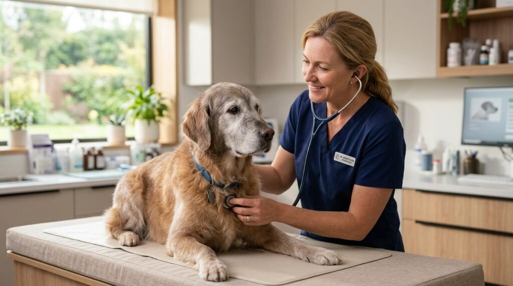
Few sounds bring as much quiet peace to a household as the rhythmic rise and fall of your senior dog resting peacefully at your feet. For years, their steady heartbeat has been the faithful background metronome of your daily life. Yet as dogs reach their senior and geriatric years, the cardiovascular system undergoes structural and mechanical changes that demand closer attention. Cardiac disease is one of the most common medical challenges faced by older canines, affecting roughly ten percent of all dogs and over sixty percent of dogs over age ten.
When you first learn that your dog has a heart murmur or notice subtle changes like a soft dry cough at night or fatigue on the evening walk, a flood of concern naturally follows. Pet parents often worry that any cardiac diagnosis signals immediate crisis or severe limitation. The reality of modern veterinary cardiology is far more hopeful. With early detection, systematic resting breath monitoring, accurate clinical staging, and targeted medications, dogs diagnosed with cardiac conditions can maintain vibrant, active lives for years.
Protecting your senior dog heart health requires transforming emotional worry into practical observation. By learning how canine heart valves and muscle chambers change with age, learning the standard veterinary staging scale, and establishing daily home monitoring habits, you become the most effective advocate your aging companion could ever have.
Quick Answer:
Maintaining senior dog heart health centers on early detection of degenerative valve disease (MMVD) or dilated cardiomyopathy (DCM) before clinical heart failure occurs. The single most vital home care habit is tracking your dog’s Sleeping Respiratory Rate (SRR) while they sleep deeply; a normal rate is under 30 breaths per minute. Key warning signs include a persistent nocturnal cough, exercise intolerance, fainting episodes (syncope), and unexplained restlessness when lying flat. Clinical diagnosis pairs stethoscopic auscultation with chest radiographs, blood pressure checks, and echocardiography to categorize the disease under standard ACVIM stages (A through D). Early pharmaceutical intervention with inodilators such as pimobendan during stage B2 significantly delays the onset of congestive heart failure and extends comfortable life expectancy.
Key Takeaways:
- High Geriatric Prevalence: More than half of small and medium breed dogs over ten years old develop some degree of valvular degeneration, usually without outward symptoms in the earliest stages.
- The 30-Breaths-Per-Minute Rule: Consistently counting your sleeping dog’s chest movements gives you the earliest detection tool for pulmonary fluid accumulation, often weeks ahead of overt distress.
- The Cardiac Cough Distinctive: A heart-related cough typically sounds dry, honking, and persistent, occurring late in the evening or early morning when the dog lies recumbent.
- Stage B2 Intervention Window: Groundbreaking clinical trials show that starting medical therapy when heart enlargement is documented on x-rays or ultrasound, even before heart failure, prolongs symptom-free life.
- Sodium and Exercise Modulation: Moderate, consistent sodium intake and gentle strolls preserve kidney-cardiac balance without placing shock stress on circulatory reserves.
The Biomechanics of the Aging Canine Heart
To grasp why senior dogs develop cardiovascular issues, it helps to review how the canine heart operates and what changes occur across a lifetime of pumping blood. The canine heart is a four-chambered muscular pump designed to move un-oxygenated blood into the lungs and propel oxygen-rich blood out to working muscles, vital organs, and the brain.
The right side of the heart receives oxygen-depleted blood from venous circulation and pumps it through the pulmonary artery into the lungs. In the pulmonary capillaries, red blood cells drop off carbon dioxide and bind fresh oxygen. This refreshed blood flows into the left atrium, passes through the one-way mitral valve into the thick muscular left ventricle, and is forcefully ejected through the aorta into systemic circulation.
As canines age, two primary pathological patterns dominate geriatric cardiovascular medicine: degenerative valvular disease and myocardial muscle disease. Recognizing which category your dog fits into depends heavily on breed size, genetics, and structural anatomy.
1. Myxomatous Mitral Valve Disease (MMVD)
By far the most common form of canine cardiac disease, Myxomatous Mitral Valve Disease accounts for roughly seventy-five to eighty percent of all heart cases evaluated in veterinary clinics. MMVD predominantly targets small to medium dog breeds, including Cavalier King Charles Spaniels, Dachshunds, Miniature Poodles, Chihuahuas, Shih Tzus, and Yorkshire Terriers, although mixed breeds and larger dogs can also be affected.
In a healthy young heart, the mitral valve leaflets are thin, flexible, translucent sheets of tissue that snap tightly shut during ventricular contraction, preventing backward blood flow into the atrium. In MMVD, a slow progressive breakdown of structural collagen and elastin occurs within the valve leaflets, replaced by mucopolysaccharides and glycosaminoglycans. The valve leaflets gradually become thickened, nodular, curled, and deformed.
Because the deformed leaflets can no longer form a tight biological seal, a portion of the blood leaks backward into the left atrium with every single beat. This backward leak is known as mitral regurgitation. The physical turbulence of this leaking jet is what your veterinarian hears through a stethoscope as a systolic heart murmur.
Over months and years, the heart must work harder to maintain systemic output. The left atrium stretches and dilates to hold the regurgitant volume. Eventually, pressure backs up through the pulmonary veins into the delicate capillary beds of the lungs, setting the stage for fluid leakage into air sacs, a condition known as pulmonary edema or left-sided congestive heart failure.
2. Dilated Cardiomyopathy (DCM)
While small dogs suffer predominantly from valve failure, large and giant breeds are more prone to primary muscle disease. Dilated Cardiomyopathy is a disease of the myocardium itself, frequently affecting Doberman Pinschers, Boxers, Great Danes, Irish Wolfhounds, Golden Retrievers, and Saint Bernards.
In DCM, the heart muscle cells lose their ability to contract forcefully. The muscular walls of the left ventricle thin out and stretch, transforming the ventricle from a strong, muscular conical shape into a thin, dilated, flaccid balloon. Because the heart muscle cannot contract strongly (a condition called systolic dysfunction), cardiac output drops drastically.
DCM is notoriously stealthy. Many dogs experience a lengthy preclinical phase with no audible murmur and no external symptoms, during which dangerous electrical arrhythmias, such as ventricular premature complexes, can trigger sudden weakness or collapse. Secondary mitral valve leakage often develops simply because the ventricle stretches so wide that the valve apparatus pulls apart.
3. Pericardial Effusion and Arrhythmias
Less common but equally critical in senior canines are conditions involving the protective sac surrounding the heart (pericardial effusion) and electrical rhythm disruptions. In older dogs, fluid can accumulate rapidly inside the fibrous pericardial sac due to benign idiopathic inflammation or vascular tumors such as hemangiosarcoma or chemodectoma. Because the fibrous sac cannot stretch quickly, rising fluid pressure compresses the heart chambers, preventing them from filling with incoming blood. This life-threatening state, known as cardiac tamponade, requires immediate veterinary pericardiocentesis to drain the trapped fluid.
Electrical rhythm disturbances (arrhythmias) can arise independently or alongside MMVD and DCM. In older dogs, aging conduction pathways can generate atrial fibrillation (a chaotic, rapid heart rate common in large breeds) or sick sinus syndrome (erratic pauses and severe slow heart rates, common in Miniature Schnauzers). Both conditions compromise blood circulation and require targeted anti-arrhythmic medications or pacemaker implantation.
Recognizing the Warning Signs: From Early Clues to Overt Symptoms
One of the greatest hurdles in senior canine healthcare is that dogs are instinctively stoic creatures. In wild ancestors, displaying weakness or respiratory vulnerability invited predation. Consequently, domestic dogs compensate for diminishing cardiac efficiency for months or even years before obvious distress surfaces. Being able to spot early, subtle behavioral signals allows you to intervene long before clinical heart failure takes hold.
Subtle Early Warning Signs
During the early phases of cardiac compensation, symptoms are often mistaken for normal aging, laziness, or mild arthritis. Watch for these initial shifts:
- Subtle Exercise Reluctance: Your dog may walk fine for five minutes, but abruptly sit down, lag behind on the leash, or refuse to complete their usual walking route.
- Delayed Recovery After Exertion: After mild activity or greeting visitors, your dog pants for fifteen to thirty minutes instead of recovering within a few minutes.
- Unexplained Restlessness at Night: When a dog lies completely flat on their side, pressure from an enlarged left atrium or early fluid shifts can make breathing slightly harder. You may notice your dog pacing, shifting sleeping spots repeatedly, or preferring to sleep with their head elevated on an armrest or pillow.
- Mild Muscle Loss Over the Back: Cardiac cachexia is a gradual wasting of lean muscle mass along the spine and thighs caused by inflammatory cytokines and altered metabolism as the heart works overtime.
- Slightly Reduced Appetite: Decreased blood flow to the digestive tract can cause mild nausea or premature fullness after eating small amounts.
Advanced and Emergent Clinical Symptoms
When the heart can no longer compensate for structural regurgitation or weak pumping, pressure spikes and clinical congestive heart failure (CHF) manifests. These signs require urgent veterinary evaluation:
- The Nocturnal Cardiac Cough: A distinctive, repetitive cough that sounds like dry hacking or throat clearing, often ending with a retching sound as if trying to expel hair or phlegm. It occurs predominantly during the night, early morning, or immediately upon standing after prolonged rest.
- Elevated Resting Respiratory Rate: Rapid, shallow chest movements during deep sleep exceeding thirty breaths per minute. For deeper insights on breathing shifts, consult our detailed analysis on old dog heavy breathing, panting causes, and when to seek urgent care.
- Orthopneic Posture: A dog struggling to breathe will sit or stand with elbows abducted (pushed outward away from the ribs) and neck extended straight forward, trying to maximize airway diameter.
- Abdominal Enlargement (Ascites): When the right side of the heart fails, fluid backs up into the abdomen, causing the belly to look distended, bloated, or pear-shaped while the spine remains bony and thin.
- Pale, Muddy, or Cyanotic Gums: Instead of healthy salmon-pink gums, poor perfusion turns oral tissues pale white, dull grayish-purple, or dusky blue, indicating severe oxygen starvation.
- Syncope (Cardiac Fainting): Sudden collapse where the dog goes limp or experiences brief stiffening of the limbs, often following excitement, coughing, or barking. Syncope occurs when the brain suffers temporary oxygen deprivation due to a sudden drop in cardiac output or an arrhythmia. Dogs typically regain consciousness within ten to sixty seconds without the prolonged disorientation seen in epileptic seizures.
The Nocturnal Cardiac Cough Versus Tracheal Collapse
A common diagnostic dilemma for owners of small senior dogs is distinguishing a cardiac cough from common respiratory ailments such as tracheal collapse or chronic bronchitis. Small breeds like Pomeranians, Yorkies, and Chihuahuas are prone to both collapsing airways and mitral valve disease, and both conditions produce persistent coughing.
A cardiac cough in dogs develops through two separate anatomical pathways:
- Left Atrial Compression: As mitral regurgitation worsens, the left atrium stretches and balloons upward. In canine anatomy, the main left bronchus (the large airway carrying air into the left lung) runs directly above the left atrium. An enlarged atrium presses upward against this sensitive bronchus, causing mechanical irritation that triggers a chronic, dry, persistent reflex cough even before any fluid enters the lungs.
- Alveolar Pulmonary Edema: When pressure in pulmonary veins exceeds the oncotic pressure of the blood, fluid leaks through capillary walls into the alveoli. This fluid irritation prompts an urgent, moist, bubbling cough, often producing white or pink tinged frothy liquid in critical stages.
In contrast, tracheal collapse typically produces a loud, dry, goose-honk cough triggered by excitement, pulling against a neck collar, drinking water, or inhaling cold air. It rarely worsens purely because the dog is lying recumbent at night, whereas a cardiac cough peaks during quiet nocturnal hours when venous blood shifts back into the central circulation.
The Gold Standard Home Tool: Sleeping Respiratory Rate (SRR)
If you implement only one single health monitoring protocol for your senior dog, make it the Sleeping Respiratory Rate (SRR), also referred to as the Resting Respiratory Rate (RRR). Decades of clinical cardiology research have proved that tracking breaths per minute while your dog is in uninterrupted sleep is the most reliable, sensitive, and cost-free home diagnostic tool available.
Pulmonary edema does not develop within seconds; it builds gradually over days as venous pressures mount. Long before your dog coughs, acts visibly sick, or refuses a meal, their sleeping breath rate begins to climb. By catching this elevation early, your veterinarian can adjust diuretic and inotropic medications, clearing the fluid before your dog suffers an acute breathing crisis requiring oxygen cage hospitalization.
How to Measure Sleeping Respiratory Rate Step-by-Step
- Wait for Deep, Peaceful Sleep: The measurement must be taken when your dog is completely relaxed, asleep, and not dreaming. Never measure your dog’s breath rate while they are awake, resting with eyes open, panting, or twitching during REM sleep dreams.
- Observe Chest Wall Movement: Watch your dog’s ribcage or flank. One complete breath consists of one rise of the chest (inhalation) and one fall of the chest (exhalation). Counting both the rise and fall as two separate breaths is a frequent owner error; remember that one rise plus one fall equals one breath.
- Count for Sixty Seconds: Using a stopwatch, clock, or smartphone timer, count the total number of breaths over a full 60-second window. Alternatively, you can count the breaths over 30 seconds and multiply by two, although a full minute provides superior precision.
- Log the Number Daily: Record the date, time, and breath count in a physical notebook or smartphone tracking app. Taking the measurement once daily at approximately the same time (such as late evening when the house is calm) establishes your dog’s personalized baseline.
Interpreting the Breath Numbers
- Normal Baseline (15 to 25 Breaths Per Minute): Most healthy senior dogs and well-managed cardiac patients breathe between twelve and twenty-four times per minute in deep sleep. Some deep-sleeping dogs may breathe as slowly as eight to ten times per minute.
- Borderline Zone (25 to 30 Breaths Per Minute): If your dog usually sleeps at eighteen breaths per minute and suddenly logs twenty-eight breaths per minute for two consecutive days, take notice. Recheck the rate several times over the next twenty-four hours.
- Clinical Threshold Alert (Sustained Above 30 Breaths Per Minute): A sleeping respiratory rate consistently exceeding thirty breaths per minute while in deep sleep is abnormal. It is the primary clinical signal of early fluid buildup in the lungs. Contact your veterinarian promptly to discuss a medication adjustment before clinical breathing distress develops.
- Emergency Alert (Above 40 to 50 Breaths Per Minute with Effort): If your dog’s breathing is visibly labored, rapid, or exceeds forty breaths per minute while resting, or if they cannot lie down comfortably, seek emergency veterinary care immediately.
The ACVIM Cardiac Staging Matrix
To standardize diagnosis, prognosis, and treatment protocols across veterinary medicine, the American College of Veterinary Internal Medicine (ACVIM) established an authoritative staging system for canine valvular and myocardial heart disease. This staging consensus categorizes patients from initial genetic risk all the way to end-stage refractory disease.
Knowing where your senior dog sits on this matrix provides immense clarity, eliminating confusion about why specific medications are prescribed or withheld at particular stages.
| ACVIM Stage | Clinical Definition & Heart Status | Clinical Symptoms | Standard Veterinary Management |
|---|---|---|---|
| Stage A | At high risk for developing heart disease due to breed genetics (Cavalier King Charles Spaniel, Dachshund, Doberman), but currently exhibits no murmur or structural abnormalities. | Zero symptoms; perfectly normal cardiac auscultation. | Routine wellness examinations; yearly stethoscopic checks; no medications or dietary changes indicated. |
| Stage B1 | Structural heart disease present; audible heart murmur detectable via stethoscope, but chest x-rays and echocardiograms show zero cardiac enlargement or chamber remodeling. | Zero outward symptoms; excellent exercise tolerance; normal breathing rates. | Baseline thoracic radiographs and echocardiogram; re-evaluation every six to twelve months; no medications required yet. |
| Stage B2 | Structural heart disease with significant cardiac remodeling; murmur is audible, and diagnostic imaging confirms left atrial and left ventricular enlargement meeting strict criteria (such as VHS > 10.5 and LA:Ao ratio > 1.6). | Asymptomatic or very mild exercise fatigue; zero pulmonary fluid; normal resting breath rate. | Initiation of pimobendan (Vetmedin) to delay heart failure onset; start daily home sleeping respiratory rate tracking; modest sodium moderation. |
| Stage C | Past or current clinical Congestive Heart Failure (CHF); elevated left-sided pressures have caused active pulmonary edema or right-sided ascites. | Nocturnal coughing, elevated sleeping breath rate (> 30 bpm), panting, exercise intolerance, weakness, orthopneic breathing. | Quadruple therapy protocol: pimobendan, loop diuretics (furosemide), ACE inhibitors (enalapril), and spironolactone; strict sodium regulation; close renal panel monitoring. |
| Stage D | End-stage refractory heart failure; clinical symptoms persist despite standard maximal doses of heart failure medications. | Recurrent pulmonary fluid, marked lethargy, severe muscle wasting, syncopal collapse, poor appetite. | Advanced specialist interventions; torsemide substitution; cough suppressants; sildenafil for pulmonary hypertension; dedicated hospice comfort care. |
Reviewing the formal consensus published in the ACVIM consensus guidelines on myxomatous mitral valve disease highlights one of the most critical shifts in modern veterinary cardiology: the importance of identifying Stage B2. In previous decades, veterinarians waited until a dog entered overt heart failure (Stage C) before starting medications. Today, clinical research demonstrates that identifying cardiomegaly and starting pimobendan in Stage B2 extends the asymptomatic period by an average of fifteen months, providing dogs more than a year of high quality life before symptoms ever appear.
Diagnostic Workup: How Veterinarians Evaluate the Canine Heart
When your family veterinarian detects a new heart murmur during a routine physical exam or when you report concerning breathing shifts, a methodical diagnostic workup is required to confirm the diagnosis and determine the exact ACVIM stage.
1. Auscultation and Murmur Grading
Every cardiac exam begins with careful stethoscopic auscultation. Your veterinarian listens to the heart across multiple valve areas on both sides of the chest, evaluating rhythm, heart rate, and the presence of abnormal murmurs or gallop sounds. Murmurs are graded on a universal scale from I to VI:
- Grade I: Very soft murmur heard only after several minutes of quiet listening in a completely silent room.
- Grade II: Soft murmur, but immediately audible when placing the stethoscope over the mitral valve.
- Grade III: Moderate-intensity murmur easily heard; approximately equal in volume to normal heart sounds.
- Grade IV: Loud murmur radiating widely across the chest wall, but lacking a palpable thrill.
- Grade V: Very loud murmur accompanied by a palpable precordial thrill (a physical vibration you can feel by resting your hand flat against the dog’s chest).
- Grade VI: Extremely loud murmur with a prominent thrill, audible even when the stethoscope is lifted slightly off the skin.
It is crucial to note that while murmur grade generally correlates with regurgitant jet turbulence, a louder murmur does not automatically mean a dog is closer to heart failure. A small dog can have a loud Grade IV murmur for four years in Stage B1 without ever suffering heart failure. Diagnostic imaging is essential to evaluate true internal heart size.
2. Thoracic Radiographs (Chest X-Rays)
Chest radiographs are an indispensable, accessible tool in cardiac evaluation. They provide a clear visual overview of cardiac silhouette size and allow immediate inspection of the lungs and pulmonary vessels. Using the standardized Vertebral Heart Score (VHS) system, veterinarians measure the long and short axes of the heart against thoracic vertebrae on the lateral x-ray. A VHS greater than 10.5 vertebrae typically suggests cardiac enlargement.
X-rays are also the primary tool used to differentiate airway diseases like chronic bronchitis from acute cardiogenic pulmonary edema, where fluffy, hazy fluid opacities appear in the perihilar lung fields.
3. Echocardiography (Cardiac Ultrasound)
Echocardiography performed by a board-certified veterinary cardiologist or experienced ultrasonographer is the undisputed gold standard of cardiac diagnostics. Ultrasound allows real-time visualization of the heart in motion. Using color-flow Doppler imaging, the clinician directly visualizes the backward jet of leaking blood, measures the exact thickness of valve leaflets, assesses ventricular muscle contraction strength, and calculates precise ratios such as the Left Atrium to Aorta (LA:Ao) diameter ratio.
An echocardiogram is the test that confirms if a dog with a murmur has reached Stage B2 heart enlargement, the point where medication is usually started. The Merck Veterinary Manual on canine heart disease explains how heart disease is diagnosed.
4. Blood Pressure, Biomarkers, and Electrocardiograms
Additional supportive diagnostics ensure a complete systemic picture:
- Blood Pressure Measurement: Systemic hypertension (high blood pressure) increases the resistance against which the left ventricle must pump, accelerating mitral regurgitation and stressing fragile vessels. Measuring blood pressure via a Doppler cuff ensures proper vascular balance.
- Cardiac Biomarker Assays (NT-proBNP): N-terminal pro-B-type natriuretic peptide is a hormone released by canine heart muscle cells when stretched under excessive pressure. A simple blood test measuring NT-proBNP levels helps distinguish if an emergency breathing crisis stems from primary respiratory disease or cardiac congestion.
- Electrocardiogram (ECG) and Holter Monitoring: An ECG evaluates electrical conductivity, detecting dangerous ventricular arrhythmias, atrial fibrillation, or heart blocks. For breeds prone to occult DCM like Dobermans or Boxers, wearing a 24-hour Holter vest records thousands of heartbeats during normal home activity to detect intermittent arrhythmias.
To prepare your companion for clinical testing, explore our practical recommendations in senior dog veterinary checkups, diagnostic testing, and preparation strategies.
Veterinary Cardiac Medications: The Four Pillars of Heart Failure Therapy
When heart disease advances to Stage B2 or Stage C, modern pharmaceuticals transform canine survival rates. Far from being simple palliative sedatives, cardiac medications are targeted pharmacological agents designed to support heart muscle contraction, reduce circulatory workload, and remove excess fluid.
Veterinary cardiologists rely on a combination of medications often referred to as the four pillars of congestive heart failure therapy:
1. Inodilators: Pimobendan (Vetmedin)
Pimobendan is one of the most significant pharmacological breakthroughs in veterinary history. It is classified as an inodilator because it accomplishes two distinct clinical actions simultaneously:
- Positive Inotrope: It sensitizes cardiac muscle fibers to existing intracellular calcium, allowing the heart to contract with greater force without burning extra cellular energy or increasing myocardial oxygen demand.
- Vasodilator: It inhibits phosphodiesterase III enzymes in peripheral blood vessel walls, relaxing systemic arteries and veins. This reduces afterload (the resistance the heart must pump against) and preload (the volume returning to the heart).
By making each pump stronger while simultaneously lowering the resistance in blood vessels, pimobendan decreases pressure inside the left atrium and reduces the regurgitant jet volume. As confirmed by the landmark EPIC clinical study, administering pimobendan to dogs in Stage B2 significantly prolongs the asymptomatic period before congestive heart failure develops.
2. Loop Diuretics: Furosemide (Salix) and Torsemide
When increased left-sided pressures push fluid into the lung tissues (Stage C pulmonary edema), diuretics are essential to save life. Furosemide acts directly on the Loop of Henle in the kidneys, blocking the reabsorption of sodium and chloride. Water follows sodium into the urine, causing the dog to produce copious amounts of dilute urine within hours of administration.
This rapid reduction in circulating intravascular blood volume pulls edema fluid out of the pulmonary air sacs and back into circulation, allowing the dog to breathe comfortably again. In advanced cases (Stage D) where dogs develop resistance to furosemide, cardiologists often substitute torsemide, a potent, longer-acting loop diuretic with enhanced bioavailability.
3. ACE Inhibitors: Enalapril and Benazepril
When cardiac output declines, the canine body mistakenly interprets the drop in arterial pressure as severe dehydration or blood loss. In response, the body activates the Renin-Angiotensin-Aldosterone System (RAAS), producing hormones that constrict blood vessels and retain sodium. While helpful during traumatic hemorrhage, this hormonal cascade is catastrophic in heart disease, forcing an already struggling heart to pump against tightly constricted vessels.
Angiotensin-Converting Enzyme (ACE) inhibitors, such as enalapril and benazepril, block the enzyme that converts angiotensin I to angiotensin II. This relaxes peripheral blood vessels, lowers systemic vascular resistance, and blunts long-term cardiac remodeling and fibrosis.
4. Aldosterone Receptor Antagonists: Spironolactone
Even with ACE inhibitor therapy, the body frequently finds alternative biological pathways to release aldosterone, an undesirable phenomenon known as aldosterone breakthrough. Aldosterone promotes potassium loss, sodium retention, and destructive myocardial fibrosis (scar tissue formation in the heart muscle). Spironolactone acts as a competitive aldosterone receptor antagonist, protecting heart tissue from fibrosis while providing gentle potassium-sparing diuretic support.
For additional clinical insights on medical heart failure management, read the clinical overview from VCA Animal Hospitals congestive heart failure clinical overview.
Daily Home Management and Supportive Care Routines
While veterinary medications manage internal hemodynamics, your day-to-day care routines at home determine your dog’s ongoing comfort, vitality, and emotional well-being. Living with a cardiac patient requires subtle adjustments to nutrition, physical exercise, and home environments.
1. Thoughtful Dietary Strategies: Sodium and Nutrients
Dietary management for senior dogs with heart disease focuses on maintaining lean body mass while avoiding excessive mineral loads that strain circulation.
- Sensible Sodium Moderation: In the past, strict sodium restriction was recommended for all dogs with heart murmurs. Modern veterinary cardiology cautions against severe sodium restriction in early stages (Stage B1 and early B2) because abrupt sodium deprivation can prematurely trigger the RAAS cascade and impair renal blood flow. In Stage C, moderate sodium restriction is warranted. The most important rule for pet parents is eliminating hidden high-sodium table scraps, such as deli meats, cheese slices, hot dogs, canned soups, and commercial rawhides soaked in brine.
- Preserving Lean Muscle: Dogs with advanced heart conditions are vulnerable to cardiac cachexia. Ensure your dog receives highly digestible, high-quality animal protein to preserve skeletal muscle strength.
- Taurine and L-Carnitine: These amino acids are vital for myocardial energy production. While essential for breeds susceptible to DCM, ensuring adequate dietary amino acids benefits all senior canines. Review dietary pros and cons in our detailed guide on grain-free senior dog food pros, cons, and nutritional balance.
- Omega-3 Fatty Acids (EPA and DHA): High-potency marine fish oil supplements provide potent anti-inflammatory benefits that suppress cachexia-inducing cytokines, improve cardiac appetite, and stabilize heart cell membranes against arrhythmias. Consult your veterinarian for appropriate weight-based dosing.
2. Exercise Modification: The Art of the Sniff Walk
A diagnosis of heart disease does not mean your senior dog must spend the rest of their days confined to a bed. Inactivity leads to muscle atrophy, joint stiffness, and depression. However, intense aerobic exercise, such as chasing balls, running alongside bicycles, or strenuous uphill hiking, must be replaced with low-impact physical activity.
Shift your daily routine toward leisurely, flat-surface sniff walks. Allow your dog to wander at their own relaxed pace, sniffing grass, hydrants, and tree trunks. Sniffing provides immense cognitive stimulation and stress reduction without raising the heart rate to dangerous aerobic thresholds. If your dog stops, sits, or pants during the stroll, pause immediately, offer water, and head home.
3. Temperature and Humidity Control
Dogs cannot sweat through their skin; their primary mechanism for shedding excess body heat is evaporative cooling through panting. For a dog with compromised heart function, vigorous panting represents intense physical labor that drives up heart rates and respiratory demands.
During hot or humid summer months, schedule outdoor bathroom breaks strictly during the cool early morning or late evening hours. Keep indoor living spaces climate-controlled with air conditioning or fans. During cold winter spells, dress your dog in an insulated sweater to prevent peripheral vasoconstriction that forces the heart to pump against stiffened blood vessels.
4. The Medication Consistency Protocol
Cardiac medications like pimobendan and furosemide operate with relatively short half-lives in the canine body. Inconsistent dosing or delayed administration creates hemodynamic peaks and valleys that can destabilize a fragile heart. Administering medications at the exact twelve-hour intervals recommended by your veterinarian ensures steady plasma concentrations and consistent cardiac support.
Keep a physical medication checklist on your refrigerator and use pill organizers to avoid accidental missed or double doses. When administering pimobendan, ensure it is given roughly one hour before meals, as food significantly impairs its oral bioavailability.
Monitoring the Kidney-Heart Connection (Cardiorenal Syndrome)
Managing senior dog heart health requires balancing the needs of two vital organ systems: the heart and the kidneys. In veterinary medicine, this intimate physiological interdependence is known as cardiorenal syndrome.
The medications that save a dog’s life from pulmonary edema (diuretics like furosemide) work by eliminating body water through the kidneys. However, if diuretic doses are too high or if the dog does not drink enough water, circulating blood volume drops, leading to pre-renal azotemia (elevated BUN and creatinine levels in blood work) and kidney distress. Conversely, if diuretics are cut back too far to spare the kidneys, fluid rapidly floods back into the lungs.
Because of this delicate balance, regular blood chemistry panels and urinalysis are mandatory for dogs receiving cardiac medications. Expect your veterinarian to recheck kidney values, blood urea nitrogen, creatinine, and electrolytes (sodium and potassium) five to seven days after any medication adjustment, and every three to four months during stable maintenance periods.
Never restrict your dog’s access to fresh drinking water while they are taking diuretic medications. Restricting water can trigger rapid, life-threatening dehydration and acute kidney injury within twenty-four hours.
Recognizing Discomfort and Evaluating Quality of Life
Heart disease can be emotionally taxing for both dog and owner. While canine cardiac disease is not physically painful in the manner of bone cancer or severe osteoarthritis, respiratory distress causes profound anxiety and discomfort. Air hunger is an intensely distressing sensation for any living creature.
Stay attuned to physical indicators of stress, restlessness, and discomfort. To help interpret subtle behavioral changes in older companions, consult our resource on senior dog pain signs, vocalization, posture, and subtle symptoms. Likewise, learning to distinguish healthy rest from exhaustion is covered in old dog sleeping patterns, normal rest, and pathological lethargy.
Use structured quality-of-life tools, such as the HHHHHMM scale (Hurt, Hunger, Hydration, Hygiene, Happiness, Mobility, More Good Days Than Bad), to objectively evaluate your dog’s daily well-being. When medical therapies reach their limits in Stage D and medications can no longer prevent persistent breathing struggles or frequent fainting spells, compassionate hospice care or peaceful euthanasia represents the final, loving gift you can provide to spare your faithful friend from suffering.
Frequently Asked Questions About Senior Dog Heart Health
Can a senior dog live a normal life with a heart murmur?
Yes, absolutely. Many senior dogs live full, joyful, and completely normal lifespans with a heart murmur. A heart murmur is simply the sound of turbulent blood flow; it is not a death sentence. Many dogs remain in ACVIM Stage B1 for years without ever experiencing heart enlargement or requiring medication. The key to long-term success is regular veterinary monitoring, annual chest x-rays or ultrasounds, and starting medical support if the heart begins to remodel.
What is the difference between heart disease and heart failure in dogs?
Heart disease refers to any structural or functional abnormality of the heart, such as a leaky mitral valve (MMVD), thickened leaflets, or weakened heart muscle (DCM). A dog can live happily with heart disease for years without feeling sick. Heart failure, specifically Congestive Heart Failure (CHF), is the clinical syndrome that occurs when diseased heart chambers can no longer pump blood efficiently, causing fluid to back up into the lungs (pulmonary edema) or abdomen (ascites). Heart disease is the underlying structural condition; heart failure is the clinical congestion that develops if disease progresses.
Why does my dog cough more at night or early in the morning?
A cardiac cough peaks at night and early in the morning for two anatomical reasons. First, when a dog lies flat on their side, venous blood pools centrally, slightly increasing pressure inside the left atrium. This stretches the atrium, which presses directly upward against the main left bronchus, triggering a mechanical cough reflex. Second, lower body temperatures, relaxed airways, and early fluid shifts during sleep increase mucosal sensitivity in the bronchial tree.
Is anesthesia safe for an older dog with a heart murmur?
Anesthesia is significantly safer today than in the past, but it requires thorough pre-operative cardiac staging. A dog in Stage B1 with a normal-sized heart handles modern, individualized anesthesia protocols with minimal elevated risk. However, a dog in Stage B2 or Stage C requires careful pre-anesthetic stabilization, customized anesthetic drugs that do not depress myocardial contractility or drop blood pressure, intravenous fluid regulation, and continuous multi-parameter monitoring by a skilled veterinary technician.
How long can a senior dog live after being diagnosed with congestive heart failure?
Survival times vary widely based on individual breed biology, age, renal resilience, and how quickly medical therapy is started. With the modern quadruple therapy protocol (pimobendan, furosemide, ACE inhibitors, and spironolactone) and vigilant home sleeping respiratory rate tracking, many dogs live comfortably for nine to eighteen months or even longer following their initial Stage C congestive heart failure diagnosis. Diligent home monitoring and swift medication adjustments when breathing rates rise are the biggest determinants of long-term longevity.